• General
  • Distribution
  • Impact
  • Management
  • Bibliography
  • Contact
Common name
pest (German), bubonic plague (English), chuma (Russian), plague (English), peste (French)
Synonym
Bacterium pestis , Lehmann and Neumann 1896
Bacillus pestis , (Lehmann and Neumann 1896) Migula 1900
Pasteurella pestis , (Lehmann and Neumann 1896) Bergey et al. 1923
Pestisella pestis , (Lehmann and Neumann 1896) Dorofeev 1947
Bacillus pestis
Bacterium pestis ,
Pasteurella pestis
Pestisella pestis ,
Similar species
Alcaligenes spp., Pseudomonas spp., Yersinia pseudotuberculosis
Summary
Yersinia pestis is a gram-negative bacterium that causes plague, a highly contagious and lethal disease and the cause of three disease pandemics throughout human history. It is a zoonotic disease and exists in natural cycles involving transmission between rodent hosts and flea vectors. Humans are usually infected through bites from rodent fleas that carry the disease. Modern antibiotics are effective against Y. pestis, but if treatment is delayed or inadequate then the disease can cause severe illness or death.
Species Description
Among the Enterobacteriaceae, Yersinia pestis is unique in both its choice of host habitat (blood, lymphoid system, reticuloendothelial system) and primary mode of transmission (flea vectors). Y. pestis has two main habitats—in the stomach of proventriculus of various flea species at ambient temperature or in the blood or tissues of a rodent host at body temperature (Perry 19997 in Prentice and Rahalison 2007). Y. pestis has been recorded to naturally infect over 203 rodent species and 14 lagomorph species. However only a small proportion are actually significant hosts, with rodents being far more important host taxa than lagomorphs (Gage and Kosoy 2005).
Notes
Curson et al. (1989) report that bubonic plague exhibits marked seasonality, closely related to seasonal changes in flea and rat populations. It tends to be a disease of late summer and early autumn, although it is possible that the artificially maintained indoor temperatures of many houses in the winter may allow fleas to survive. It is also possible that Y. pestis can be transmitted to humans via lice and other insects.
Fleas require fairly specific climatic conditions and do best in moderately warm (15-20 degrees Celsius) and moist climates (90-95% humidity). In species of fleas such as Xenopsylla cheopis , Y. pestis reproduces most rapidly at warm temperatures of up to 27 degrees Celsius. Above this temperature, changes occur in blood coagulation, resulting in digestion of the blocking plug and the rapid elimination of Y. pestis. Y. pestis produces two antiphagocytic components (that impede or prevent the function of defensive white blood cells), called F1 capsule antigen and the V antigen. Both are required for the disease to be harmful and are only produced when the host's body temperature is not lower than 37 degrees Celsius (Fix 1997).
Lifecycle Stages
Yersinia pestis is maintained in nature through transmission between hematophagous [blood feeding] adult fleas and certain rodent hosts, with occasional involvement of some lagomorphs (Gage 1998; Pollitzer 1961)” (Gage and Kosoy 2005). Evidence of Y. pestis infection in a wide range of other mammal groups suggests that virtually all mammals are susceptible to this bacterium (Gage 1998; Gage 1999; Pollitzer 1961 in Gage and Kosoy 2005).

“Typically, plague is thought to exist indefinitely in so-called enzootic (maintenance) cycles that cause little obvious host mortality and involve transmission between partially resistant rodents (enzootic or maintenance hosts) and their fleas (Gage et al 1995; Poland and Barnes 1979; Poland et al. 1994). Occasionally, the disease spreads from enzootic hosts to more highly susceptible animals, termed epizootic or amplifying hosts, often causing rapidly spreading die-offs (epizootics)” (Gage and Kosoy 2005). Humans and other highly susceptible mammals also experience their greatest exposure risks during epizootics.”

However, there is some debate over whether epizootic and enzootic cycles actually exist. In North America plague causes die-offs of colonies of prairie dogs (Cynomys ludovicianus). “It has been argued that other small rodents are reservoirs for plague, spreading disease during epizootics and maintaining the pathogen in the absence of prairie dogs; yet there is little empirical support for distinct enzootic and epizootic cycles.” Stapp et al. (2008) investigated a number of small rodent species in northern Colorado, and found no evidence that any small rodent acts as a long-term, enzootic host for Y. pestis in prairie dog colonies.

The question of whether Y. pestis can survive outside its normal host or vector has been a controversial issue. A recent study by Ayyadurai et al. (2008) confirmed that Y. pestis remains viable and virulent after 40 weeks incubation in sterilized humidified sand. Survival in soil is clearly an important mechanism for plague persistence during inter-epizootic periods and plays an important role in the epidemiology of the plague (Ayyadurai et al. 2008).

The main vectors responsible for transmission of Y. pestis to humans are usually rodent fleas, Xenopsylla cheopis and Nosopsylla fasciatus, or in some cases the human flea, Pulex irritans (Curson et al. 1989). In North America the primary vector of Y. pestis to humans is Oropsylla Montana (Eisen et al. 2007-C). When a flea bites its host it ingests Y. pestis and becomes infected. Y. pestis may reproduce so rapidly that it blocks the flea's proventriculus, a small organ located between the esophageus and stomach. This block prevents any ingested blood from reaching the midgut, causing the flea to starve. Regurgitation of ingested blood and infectious material from the blockage are forced back into the wound, infecting the host. This combined with increased feeding attempts from starvation make blocked fleas dangerous vectors of Y. pestis (Eisen et al. 2007-D). Spread by blocked fleas has been the accepted paradigm for plague transmission for many years.

However Eisen et al. (2007-D) point out “that this mechanism, which requires a lengthy extrinsic incubation period before a short infectious window often followed by death of the flea, cannot sufficiently explain the rapid rate of spread that typifies plague epidemics and epizootics” and explain the importance of unblocked fleas in Y. pestis epizootics. Unblocked fleas are immediately infectious, transmit the bacterium for at least 4 days, and remain infectious for long periods as they do not suffer block-induced mortality.

Uses
Yersinia pestis has the potential to be used as a weapon in bioterrorism. The United States Centers for Disease Control classified Y. pestis a Category A Select Agent due to its potential to pose a severe threat to public health and safety. Concerns stem from the fact that during the Cold War, both American and Soviet scientists devised means to effectively aerosolize Y. pestis, thereby removing the need for the flea vector (Inglesby 2000 in Smiley 2008). Although antibiotics are effective against plague, antibiotic resistant strains are known to exist. \"Covertly aerosolized, antibioticresistant Y. pestis would be a formidable weapon of terror\" (Smiley 2008).
Habitat Description
Among the Enterobacteriaceae, Yersinia pestis is unique in both its choice of host habitat (blood, lymphoid system, reticuloendothelial system) and primary mode of transmission (flea vectors). Y. pestis has two main habitats—in the stomach of proventriculus of various flea species at ambient temperature or in the blood or tissues of a rodent host at body temperature (Perry 19997 in Prentice and Rahalison 2007). Y. pestis has been recorded to naturally infect over 203 rodent species and 14 lagomorph species. However only a small proportion are actually significant hosts, with rodents being far more important host taxa than lagomorphs (Gage and Kosoy 2005).
Reproduction
According to Campbell et al. (1999), bacteria reproduce asexually using binary fission. Binary fission is a type of cellular division in which each dividing daughter cell receives a copy of the single parent chromosome. Growth of bacteria can be extremely fast if the resources needed are not limited and the colonies do not poison themselves with the accumulation of their own wastes (Campbell et al. 1999). The generation time (the time it takes for the colony to double in size) is 1.25 hours (Chu 2001).
Nutrition
Campbell et al. (1999) write that in order to grow in nature or in the laboratory, a bacterium must have an energy source, a source of carbon and other required nutrients, and a permissive range of physical conditions such as oxygen concentration, temperature, and pH. Y. pestis is a chemoheterotroph, meaning that it must consume organic molecules for energy and carbon.
Pathway
The bacteria live in fleas, which are carried by rats, rabbits, humans and other mammals. These animals can be transported around the world with human cargo. Humans can carry the fleas and the disease unknowingly during the 1 to 6 day incubation period.

Principal source: Gage, K. L., D. T. Dennis, and T. F. Tsai. 2001. Prevention of Plague: Recommendations of the Advisory Committee on Immunization Practices (ACIP). Center for Disease Control, Morbidity and Mortality Weekly Report.

Compiler: National Biological Information Infrastructure (NBII) & IUCN/SSC Invasive Species Specialist Group (ISSG)
Updates completed with support from the Ministry of Agriculture and Forestry (MAF)- Biosecurity New Zealand

Review: Dr James B. Bliska \ Department of Molecular Genetics and Microbiology \ Center for Infectious Diseases \ Stony Brook, NY USA.

Publication date: 2006-03-31

Recommended citation: Global Invasive Species Database (2024) Species profile: Yersinia pestis. Downloaded from http://www.iucngisd.org/gisd/species.php?sc=450 on 20-04-2024.

General Impacts
Yersinia pestis is the causal agent of plague in humans and other mammals, although the overwhelming proportion of attention and research has focused on its impacts on humans. Y. pestis is recognized as causing three major disease pandemics in the 1st, 14th-17th and 19th centuries, resulting in around 200 million deaths. The second pandemic known as the Black Death caused the deaths of over 30% of the population of Europe. While Y. pestis no longer causes problems of such magnitude, it is still a public health concern in Africa, Asia and South America (Titball and Williamson 2001). There are at least 2000 cases of plague reported annually. In the United States it is a rare disease of humans, with only 112 cases reported between 1988-2002, although fatality rates remain high (MNWR 2002 in Eisen et al. 2007-B).

Biologists are increasingly realizing that wild mammal species are highly susceptible to Y. pestis. In North America more than half of rodent species of conservation concern occur within the range of Y. pestis. The impacts of plague on these populations are not well understood, but certain features increase the vulnerability of rodent species to plague. These include low natural resistance, high population densities, coloniality and sociality, abundant flea vectors, and lack of ability to cope with high demographic or environmental stochasticity.

Please follow this link for more details on the impacts of Yersinia pestis.

Management Info
Please follow this link for more details on the prevention, management and control of the spread of Yersinia pestis
Countries (or multi-country features) with distribution records for Yersinia pestis
Informations on Yersinia pestis has been recorded for the following locations. Click on the name for additional informations.
Lorem Ipsum
Location Status Invasiveness Occurrence Source
Details of Yersinia pestis in information
Status
Invasiveness
Arrival date
Occurrence
Source
Introduction
Species notes for this location
Location note
Management notes for this location
Impact
Mechanism:
Outcome:
Ecosystem services:
Impact information
Yersinia pestis is the causal agent of plague in humans and other mammals, although the overwhelming proportion of attention and research has focused on its impacts on humans. Y. pestis is recognized as causing three major disease pandemics in the 1st, 14th-17th and 19th centuries, resulting in around 200 million deaths. The second pandemic known as the Black Death caused the deaths of over 30% of the population of Europe. While Y. pestis no longer causes problems of such magnitude, it is still a public health concern in Africa, Asia and South America (Titball and Williamson 2001). There are at least 2000 cases of plague reported annually. In the United States it is a rare disease of humans, with only 112 cases reported between 1988-2002, although fatality rates remain high (MNWR 2002 in Eisen et al. 2007-B).

Biologists are increasingly realizing that wild mammal species are highly susceptible to Y. pestis. In North America more than half of rodent species of conservation concern occur within the range of Y. pestis. The impacts of plague on these populations are not well understood, but certain features increase the vulnerability of rodent species to plague. These include low natural resistance, high population densities, coloniality and sociality, abundant flea vectors, and lack of ability to cope with high demographic or environmental stochasticity.

Please follow this link for more details on the impacts of Yersinia pestis.

Outcomes
[7] Environmental Species - Population
  • [7] Plant/animal health
[4] Socio-Economic
  • [4] Human health
Management information
Please follow this link for more details on the prevention, management and control of the spread of Yersinia pestis
Locations
PERU
UGANDA
UNITED STATES
Management Category
Control
Unknown
Bibliography
63 references found for Yersinia pestis

Management information
Anisimov, A.P. & Amoako, K. 2006. Treatment of plague: promising alternatives to antibiotics. Journal of Medical Microbiology 55: 1461-1475.
Blisnick, T., Ave, P., Huerre, M., Carniel, E. & Demeure, C.E. 2008. Oral vaccination against bubonic plague using a live avirulent Yersinia pseudotuberculosis strain. Infection and Immunity 76(8): 3808-3816.
Center for Disease Control. February 25, 2002. Plague . Center for Disease Control.
Summary: Brief report on symptoms, description, global distribution, control and preventive measures.
Available from: http://www2.ncid.cdc.gov/travel/yb/utils/ybGet.asp?section=dis&obj=plague.htm [Accessed 25 June 2006].
Center for Disease Control. February 25, 2002. Plague. Center for Disease Control
Summary: Brief report on symptoms, description, global distribution, control and preventive measures.
Available from: http://www2.ncid.cdc.gov/travel/yb/utils/ybGet.asp?section=dis&obj=plague.htm [Accessed 25 June 2006].
Chu, May C. April 14, 2001. Basic Laboratory Protocols For The Presumptive Identification of Yersinia pestis . Center For Disease Control.
Summary: Detailed report on history, impacts, control and treatment, description, and protocols for laboratory procedure when dealing with Yersinia pestis.
Cornelius, C.A., Quenee, L.E., Overheim, K.A., Koster, F., Brasel, T.L., Elli, D., Ciletti, N.A. & Schneewind, O. 2008. Immunization with recombinant V10 protects cynomolgus macaques from lethal pneumonic plague. Infection and Immunity 76(12): 5588-5597.
Fix, D. 1997. Yersinia. Center for Environmental Health Web Server at Southern Illinois University at Carbondale
Summary: Report on description, general impacts, and control and preventive measures used.
Available from: http://www.cehs.siu.edu/fix/medmicro/yersi.htm [Accessed 17 February 2003].
Fix, Douglas. 1997. Yersinia . Center for Enviromental Health Web Server at Southern Illinois University at Carbondale
Summary: Report on description, general impacts, and control and preventive measures used.
Available from: http://www.cehs.siu.edu/fix/medmicro/yersi.htm [Accessed 17 February 2003].
Gage, K.L., Dennis, D.T. & Tsai, T.F. 2001. Prevention of Plague: Recommendations of the Advisory Committee on Immunization Practices (ACIP). Center for Disease Control, Morbidity and Mortality Weekly Report.
Summary: Detailed report on symptoms, hosts, human exposure risks, and possible control and preventive measures.
Available from: http://www.cehs.siu.edu/fix/medmicro/yersi.htm [Accessed 17 February 2003].
Kummer, L.W., Szaba, F.M., Parent, M.A., Adamomvicz, J.J., Hill, J., Johnson, L.L. & Smiley, S.T. 2008. Antibodies and cytokines independently protect against pneumonic plague. Vaccine 26: 6901-6907.
Morris, S.R. 2007. Development of a recombinant vaccine against aerosolized plague. Vaccine 25: 3115-3117.
Rocke, T.E., Smith, S., Marinari, P., Kreeger, J., Enama, J.T. & Powell, B.S. 2008-B. Vaccination with F1-V fusion protein protects black-footed ferrets (Mustela nigripes) against plague upon oral challenge with Yersinia pestis. Journal of Wildlife Diseases 44(1): 1-7.
Rocke, T.E., Smith, S.R., Stinchcomb, D.T. & Osorio, J.E. 2008-A. Immunization of black-tailed prairie dogs against plague through consumption of vaccine-laden baits. Journal of Wildlife Diseases 44(4): 930-937.
Seery, D.B., Biggins, D.E., Montenieri, J.A., Enscore, R.E., Tanda, D.T. & Gage, K.L. 2003. Treatment of black-tailed prairie dog burrows with Deltamethrin to control fleas (Insecta: Siphonaptera) and plague. Journal of Medical Entomology 40(5): 718-722.
Smiley, S.T. 2008. Immune defense against pneumonic plague. Immunological Reviews 225: 256-271.
Titball, R.W. & Williamson, E.D. 2001. Vaccination against bubonic and pneumonic plague. Vaccine 19: 4175-4184.
General information
Adjemian, J.Z., Foley, P., Gage, K.L. & Foley, J.E. 2007. Initiation and spread of traveling waves of plague, Yersinia pestis, in the western United States. American Journal of Tropical Medicine and Hygiene 76(2): 365-375.
Anisimov, A.P., Lindler, L.E. & Pier, G.B. 2004. Intraspecific diversity of Yersinia pestis. Clinical Microbiology Reviews 17(2): 434-464.
Augustine, D.J., Dinsmore, S.J., Wunder, M.B., Dreitz, V.J & Knopf, F.L. 2008. Response of mountain plovers to plague-driven dynamics of black-tailed prairie dog colonies. Landscape Ecology 23: 689-697.
Ayyadurai, S., Houhamdi, L., Lepidi, H., Nappez, C., Raoult, D. & Drancourt, M. 2008. Long-term persistence of virulent Yersinia pestis in soil. Microbiology 154: 2865-2871.
Bertherat, E., Bekhoucha, S., Chougrani, S., Razik, F., Duchemin, J.B., Houti, L., Deharib, L., Favolle, C., Makrerougrass, R.D., Bella, R., Belhabri, L., Chaieb, A., Evgueni, T. & Carniel, E. 2007. Plague reappearance in Algeria after 50 years, 2003. Emerging Infectious Diseases 13(10).
Biggins, D.E. & Kosoy. 2001. Influences of introduced plague on North American mammals: implications from ecology of plague in Asia. Journal of Mammalogy 82(4): 906-916.
Campbell, N.A., Reece, J.B., Mitchell, L.G. 1999. Biology 5th Edition. Menlo Park, California: Addison Wesley Longman Inc.
Summary: A biology textbook. Used for bacteria reproduction info.
Campbell, Neil A.; Reece, Jane B.; Mitchell, Lawrence G.. 1999. Biology Fifth Edition . Addison Wesley Longman, Inc. Menlo Park, California.
Summary: A biology textbook. Used for bacteria reproduction info.
Center for Disease Control. June 22, 2001. CDC Plague Home Page . Center for Disease Control.
Summary: Brief summary of transmission and risk of exposure. Includes map of global distribution.
Available from: http://www.cdc.gov/ncidod/dvbid/plague/index.htm [Accessed 17 February 2003].
Center for Disease Control. June 22, 2001. CDC Plague Home Page. Center for Disease Control.
Summary: Brief summary of transmission and risk of exposure. Includes map of global distribution.
Available from: http://www.cdc.gov/ncidod/dvbid/plague/index.htm [Accessed 17 February 2003].
Centers for Disease Control and Prevention. 2006. Human plague � four states. [Accessed January 12, 2009]
Summary: Available from: http://www.cdc.gov/mmwr/preview/mmwrhtml/mm5534a4.htm [Accessed 25 June 2009]
Chu, M.C. 2001. Basic laboratory protocols for the presumptive identification of Yersinia pestisSummary: Detailed report on history, impacts, control and treatment, description, and protocols for laboratory procedure when dealing with Yersinia pestis.
Collinge, S.K., Johnson, W.C., Ray, C., Matchett, R., Grensten, J., Cully, J.F., Gage, K.L., Kosoy, M.Y., Loye, J.E & Martin, A.P. 2005. Testing the generality of a trophic-cascade model for plague. EcoHealth 2: 102-112.
Cui, Y., Li, Y., Gorge, O., Platonov, M.E., Yan, Y., Guo, Z., Pourcel, C., Dentovskay, S.V., Balakhonov, S.V., Wang, X., Song, Y., Anisimov, A.P., Vergnaud, G. & Yang, R. 2008. Insight into microevolution of Yersinia pestis by clustered regularly interspaced short palindromic repeats. Public Library of Science ONE 3(7).
Cully, J.F., Barnes, A.M., Quan, T.J & Maupin, G. 1997. Dynamics of plague in a Gunnison�s prairie dog colony complex from New Mexico. Journal of Wildlife Diseases 33(4): 706-719.
Curson, P. and McCracken, K.. 1989. Plague in Sydney: An Anatomy of an Epidemic. Kensington: NSW University Press.
Summary: Detailed description of vectors of transmission.
Available from: http://pandora.nla.gov.au/pan/13025/20040119/www.maps.jcu.edu.au/course/hist/fever/plague/plague.html [Accessed 17 February 2003].
Curson, P. & McCracken, K. 1989. Plague in Sydney: An Anatomy of an Epidemic. Kensington: NSW University Press.
Summary: Detailed description of vectors of transmission.
Available from: http://pandora.nla.gov.au/pan/13025/20040119/www.maps.jcu.edu.au/course/hist/fever/plague/plague.html [Accessed 17 February 2003].
Edmunds, D.R., Williams, E.S., O�Toole, D., Mills, K.W., Boerger-Fields, A.M., Jaeger, P.T., Bildfell, R.J., Dearing, P. & Cornish, T.E. 2008. Ocular plague (Yersinia pestis) in mule deer (Odocoileus hemionus) from Wyoming and Oregon. Journal of Wildlife Diseases 44(4): 983-987.
Eisen, R.J., Bearden, S.W., Wilder, A.P., Montenieri, J.A., Antolin, M.F. & Gage, K.L. 2007-D. Early-phase transmission of Yersinia pestis by unblocked fleas as a mechanism explaining rapidly spreading plague epizootics. Proceedings of the National Academy of Sciences 103(42): 1538-15385.
Eisen, R.J., Borchert, J.N., Holmes, J.L., Amatre, G., Van Wyk, K., Enscore, R.E., Babi, N, Atiku, L.A., Wilder, A.P., Vetter, S.M., Bearden, S.W., Montenieri, J.A., & Gage, K.L. 2008. Early-phase transmission of Yersinia pestis by cat fleas (Ctenocephalides felis and their potential role as vectors in a plague-endemic region of Uganda. American Journal of Tropical Medicine and Hygiene 78(6): 949-956.
Eisen, R.J., Enscore, R.E., Biggerstaff, B.J., Reynolds, P.J., Ettestad, P., Brown, T., Pape, J., Tanda, D., Levy, C.E., Engelthaler, D.M., Cheek, J., Bueno, R., Targhetta, J., Montenieri, J.A. & Gage, K.L. 2007-B. Human plague in the southwestern United States, 1957-2004: spatial models of elevated risk of human exposure to Yersinia pestis. Journal of Medical Entomology 44(3): 530-537.
Eisen, R.J., Lowell, J.L., Montenieri, J.A., Bearden, S.W. & Gage, K.L. 2007-C. Temporal dynamics of early-phase transmission of Yersinia pestis by unblocked fleas: secondary infectious feeds prolong efficient transmission by Oropsylla Montana (Siphonaptera: Ceratophyllidae). Journal of Medical Entomology 44(4): 672-677.
Eisen, R.J., Reynolds, P.J., Ettestad, P., Brown, T., Enscore, R.E., Biggerstaff, B.J., Cheek, J., Bueno, R., Targhetta, J., Montenieri, J.A., & Gage, K.L. 2007-A. Residence-linked human plague in New Mexico: a habitat-suitability model. American Journal of Tropical Medicine and Hygiene 77(1): 121-125.
Euzeby, J.P. 1997. List of bacterial names withstanding in nomenclature � Genus Yersinia. Society for Systematic and Veterinary Bacteriology. �cole Nationale V�t�rinaire de Toulouse, France.
Summary: List of current and previous full scientific names.
Available from: http://www.bacterio.cict.fr/xz/yersinia.html [Accessed 17 February 2003].
Euz�by, J.P.. 1997. List of Bacterial Names with Standing in Nomenclature - Genus Yersinia. Society for Systematic and Veterinary Bacteriology; �cole Nationale V�t�rinaire de Toulouse, France.
Summary: List of current and previous full scientific names.
Available from: http://www.bacterio.cict.fr/xz/yersinia.html [Accessed 17 February 2003].
Gage, K.L. & Kosoy, M.Y. 2005. Natural history of plague: perspectives from more than a century of research. Annual Review of Entomolgoy 50: 505-528.
Garcia, E., Worsham, P., Bearden, S., Malfatti, S., Lang, D., Larimer, F., Lindler, L. & Chain, P. 2007. Pestoides F, an atypical Yersinia pestis strain from the Former Soviet Union. In The Genus Yersina. New York: Springer.
Guarner, J., Shieh, W.J., Chu, M., Perlman, D.C., Kool, J., Gage, K.L., Ettestad, P. & Zaki, S.R. 2005. Persistent Yersinia pestis antigens in ischemic tissues of a patient with septicemic plague. Human Pathology 36: 850-853.
International Society for Infectious Diseases. 2004. Plague, feline � USA (Colorado).
International Society for Infectious Diseases. 2007. Undiagnosed deaths � Zambia (02): pneumonic plague suspected.
International Society for Infectious Diseases. 2008-A. Plague � Peru: (Lambayeque), potential for outbreak.
International Society for Infectious Diseases. 2008-B. Plague, pneumonic � Madagascar: (Toamasina).
International Society for Infectious Diseases. 2008-C. Plague, wildlife, human exposure � Russia (Siberia).
International Society for Infectious Diseases. 2008-D. Plague, fatal - Madagascar: (Antananarivo).
Liang, J.M., Li, H.Y., Zeng, J., Deng, Q.Y., Zhou, S.W., Wei, J.P., Chen, D.Z., Li, S.S., Qin, S.Y., Huang, D.H., Lu, C.F., Liao, C.Z., Fu, J.Y. & Jue, G.Y. 2007. Investigation into plague status in Longtan Reservoir region in Guangxi. Chinese Journal of Endemiology 26(5): 548-550.
Liu, H.Z., Du, G.Y., Bai, X.W., Zhang, Y.Z., Shi, X.M., Wang, H.F., Bai, X.Y, Hu, L.L. & Yang, S.L. 2007. The virulence test of Yersinia pestis from a plague foci in Hebei Province. Chinese Journal of Endemiology 26(5): 478-480.
Li, Y., Dai, E., Cui, Y., Li, M., Zhang, Y., Wu, M., Zhou, D., Guo, Z., Dai, X., Cui, B., Qi, Z., Wang, Z., Wang, H., Dong, X., Song, Z., Zhai, J., Song, Y. & Yang, R. 2008. Different region analysis for genotyping Yersinia pestis isolates from China. Public Library of Science ONE 3(5): 1-10.
Lowell, J.L., Wagner, D.M., Atshabar, B., Antolin, M.F., Vogler, A.J., Keim, P., Chu, M.C., Gage, K.L. 2005. Identifying Sources of Human Exposure to Plague. Journal of Clinical Microbiology 43(2): 650-656.
Prentice, M.B. & Rahalison, L. 2007. Plague. Lancet 369: 1196-1207.
Qi, Z.Z., Wang, Z.Y., He, J., Wei, R.J., Zhao, H.H., Zhang, Q.W., Yang, X.Y., Li, C.X., Dai, R.X., Yang, Y.H., Yin, Y.Q., Zhai, H.T., Mao, X.H., Qi, H.W. & Xiao, Y. 2008. Study on plague pathogeny in areas along Qinghai-Tibet Railroad in Qinghai from 2001 � 2006. Chinese Journal of Endemiology 27(2): 204-206.
Songer, Glenn. Undated. Yersinia pestis: The Plague through History . University of Arizona.
Summary: Lecture notes about the history of Yersinia pestis from the University of Arizona.
Songer, G. n.d. Yersinia pestis: the plague through history. University of Arizona.
Summary: Lecture notes about the history of Yersinia pestis from the University of Arizona.
Stapp, P., Salkeld, D.J., Eisen, R.J., Pappert, R., Young, J., Carter, L.G., Gage, K.L., Tripp, D.W. & Antolin, M.F. 2008. Exposure of small rodents to plague during epizootics in black-tailed prairie dogs. Journal of Wildlife Diseases 44(3): 724-730.
Stenseth, N.C., Atshabar, B.B., Begon, M., Belmain, S.R., Bertherat, E., Carniel, E., Gage, K.L., Leirs, H., Rahalison, L. 2008. Plague: past, present and future. Public Library of Science Medicine 5(1): 9-13.
Thiagarajan, B., Bai, Y., Gage, K.L. & Cully, J.F. 2008. Prevalence of Yersinia pestis in rodents and fleas associated with black-tailed prairie dogs (Cynomys ludovicianus) and thunder basin national grassland, Wyoming. Journal of Wildlife Diseases 44(3): 731-736.
Wilder, A.P., Eisen, R.J., Bearden, S.W., Montenieri, J.A., Tripp, D.W., Brinkerhoff, R.J., Gage, K.L. & Antolin, M.F. 2008. Transmission efficiency of two flea species (Oropsylla tuberculata cynomuris and Oropsylla hirsute involved in plague epizootics among prairie dogs. EcoHealth 5: 205-212.
Contact
The following 5 contacts offer information an advice on Yersinia pestis
Anisimov,
Andrey P.
Organization:
Deputy Director for Science
State Research Center for Applied Microbiology & Biotechnology,
Address:
P.O. Box 13,
142279 Obolensk,Serpukhov District,
Moscow Region,
Russia
Phone:
7-4967-36-01-17
Fax:
7-4967-36-00-61
B. Bliska,
Dr James
Pathogenic bacteria.
Organization:
Cemnter for Infectious Diseases
Address:
Department of Molecular Genetics and Microbiology SUNY Stony Brook, NY 11794-5222
Phone:
631-632-8782
Fax:
631-632-9797
Eisen,
Rebecca J.
Organization:
Centers for Disease Control and Prevention
Email:
Address:
Centers for Disease Control and Prevention, 3150 Rampart Road, Fort Collins, CO 80522
Phone:
970-221-6408
Fax:
970-221-6476
Gage,
Kenneth L.
I am chief of the Flea-Borne Diseases Activity at the U.S. Centers for Disease Control and Prevention s Division of Vector-Borne Infectious Diseases. My group and I have expertise in the epidemiology, ecology, surveillance and control of plague. We also have active research programs and numerous publications on the factors influencing transmission of Yersinia pestis by fleas, ecological and climatic factors influencing the occurrences of plague outbreaks, GIS-based modeling of human plague risks and plague foci, and the mechanisms of plague maintenance in natural foci.
Organization:
Bacterial Zoonoses Branch, Division of Vector-Borne Infectious Diseases,
Centers for Disease Control and Prevention
Email:
Address:
Fort Collins, Colorado 80523;
Phone:
970-221-6450
Fax:
970-221-6476
Yang,
Ruifu
Organization:
Beijing Institute of Microbiology and Epidemiology
Address:
No.20, Dongdajie, Fengtai District,
Beijing 100071
P.R. of China
Phone:
Fax:
0086-10-63815689
Yersinia pestis
pest, bubonic plague, chuma, plague, peste
Date assessed
Year published
Eicat category
Justification for EICAT assessment
Confidence rating
Mechanism(s) of maximum impact
Countries of most severe impact
Description of impacts
Assessor
Contributors
Reviewers
Recommended citation
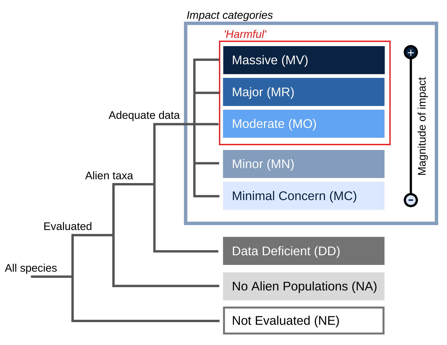
(2024). Yersinia pestis. IUCN Environmental Impact Classification for Alien Taxa (EICAT).